lv endocardium | left ventricular arrhythmia map lv endocardium The heart is comprised of the pericardium, myocardium, and endocardium. Pathology in any of those structures can lead to heart failure. Left ventricular failure occurs when there is dysfunction of the left ventricle causing insufficient delivery of blood to vital body organs. In this article, we explore the potentially destructive voltage spikes that result from the seemingly innocuous act of applying power to a circuit with a low equivalent series resistance (ESR) capacitor across its power input.
0 · lv endocardial pacing
1 · left ventricular summit arrhythmia diagram
2 · left ventricular outflow anatomy
3 · left ventricular arrhythmia map
4 · endocardial lv pacing chart
5 · endocardial left ventricular pacing
6 · endocardial electrode implantation
7 · diagram of left ventricular arrhythmia
Pērc jebkuru Cido 1l zaļo sulas paku un laimē sulīgas balvas! Vairāk informācija www.cido.lv
lv endocardial pacing
Endocardial pacing enables stimulation of the LV endocardium at any location, unrestricted by coronary venous anatomy, therefore enabling pacing at the latest activation . The heart is comprised of the pericardium, myocardium, and endocardium. Pathology in any of those structures can lead to heart failure. Left ventricular failure occurs when there is dysfunction of the left ventricle causing insufficient delivery of blood to vital body organs.
left ventricular summit arrhythmia diagram
Endocardial pacing enables stimulation of the LV endocardium at any location, unrestricted by coronary venous anatomy, therefore enabling pacing at the latest activation site and away from myocardial scar.
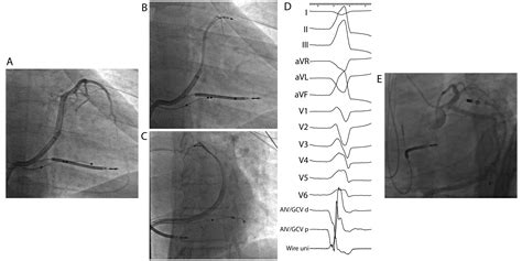
LVS VAs can be eliminated by ablation from the coronary venous system or from adjacent endocardial structures, including the LCC, basal LV endocardium, or septal RVOT.
Left ventricular (LV) endocardial pacing allows pacing at site-specific locations that enable the operator to avoid myocardial scar and target areas of latest activation. Left bundle branch area pacing (LBBAP) provides a more physiological activation pattern and may allow effective cardiac resynchronisation.
Overview. Left ventricular hypertrophy Enlarge image. Left ventricular hypertrophy is a thickening of the wall of the heart's main pumping chamber, called the left ventricle. This thickening may increase pressure within the heart. The condition can .
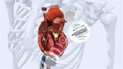
Left ventricular (LV) endocardial pacing is a promising method to deliver cardiac resynchronization therapy (CRT). WiSE-CRT is a wireless LV endocardial pacing system, and delivers ultrasonic energy to an LV electrode.Cardiac MRI(cMRI) revealed moderate left ventricle dilatation with severe dysfunction and heavy trabeculations of LV apex and mid-LV endocardium with a compact to non-compact wall ratio of less than 1:2 consistent with non-compaction cardiomyopathy .
In this review, we will describe the different techniques proposed to allow LV endocardial pacing, the results observed, and then we will discuss the reasons why LV endocardial pacing seems to be out of fashion today and what are the possible perspectives for development.
left ventricular outflow anatomy
The most common indications for endocardial LV pacing were for difficult CS anatomy (n = 12; 34%), failure to respond to conventional CRT (n = 10; 29%), and a high CS pacing threshold or phrenic nerve capture at low outputs (n = 5; 14%). From the RA side of the PSP-LV, a small atrial signal and a larger ventricular signal were recorded in each case, with an activation time of 32±7 ms pre-QRS (versus 16±5 ms pre-QRS in the LV endocardium; P=0.068). We were able to capture the LV from these sites. The heart is comprised of the pericardium, myocardium, and endocardium. Pathology in any of those structures can lead to heart failure. Left ventricular failure occurs when there is dysfunction of the left ventricle causing insufficient delivery of blood to vital body organs. Endocardial pacing enables stimulation of the LV endocardium at any location, unrestricted by coronary venous anatomy, therefore enabling pacing at the latest activation site and away from myocardial scar.

LVS VAs can be eliminated by ablation from the coronary venous system or from adjacent endocardial structures, including the LCC, basal LV endocardium, or septal RVOT. Left ventricular (LV) endocardial pacing allows pacing at site-specific locations that enable the operator to avoid myocardial scar and target areas of latest activation. Left bundle branch area pacing (LBBAP) provides a more physiological activation pattern and may allow effective cardiac resynchronisation. Overview. Left ventricular hypertrophy Enlarge image. Left ventricular hypertrophy is a thickening of the wall of the heart's main pumping chamber, called the left ventricle. This thickening may increase pressure within the heart. The condition can .
Left ventricular (LV) endocardial pacing is a promising method to deliver cardiac resynchronization therapy (CRT). WiSE-CRT is a wireless LV endocardial pacing system, and delivers ultrasonic energy to an LV electrode.
Cardiac MRI(cMRI) revealed moderate left ventricle dilatation with severe dysfunction and heavy trabeculations of LV apex and mid-LV endocardium with a compact to non-compact wall ratio of less than 1:2 consistent with non-compaction cardiomyopathy .
In this review, we will describe the different techniques proposed to allow LV endocardial pacing, the results observed, and then we will discuss the reasons why LV endocardial pacing seems to be out of fashion today and what are the possible perspectives for development.The most common indications for endocardial LV pacing were for difficult CS anatomy (n = 12; 34%), failure to respond to conventional CRT (n = 10; 29%), and a high CS pacing threshold or phrenic nerve capture at low outputs (n = 5; 14%).
saks baby gucci

left ventricular arrhythmia map
endocardial lv pacing chart
endocardial left ventricular pacing
1. Look them up on the official jail inmate roster. 2. Look them up on vinelink.com, a national inmate tracking resource. 3. Call the jail at 702-229-6444. This is available 24 hours a day. 4. Write or visit the jail and request information. You can reach them at: Las Vegas Detention Center. 3200 Stewart Avenue. Las Vegas, NV 89101. 5.
lv endocardium|left ventricular arrhythmia map


